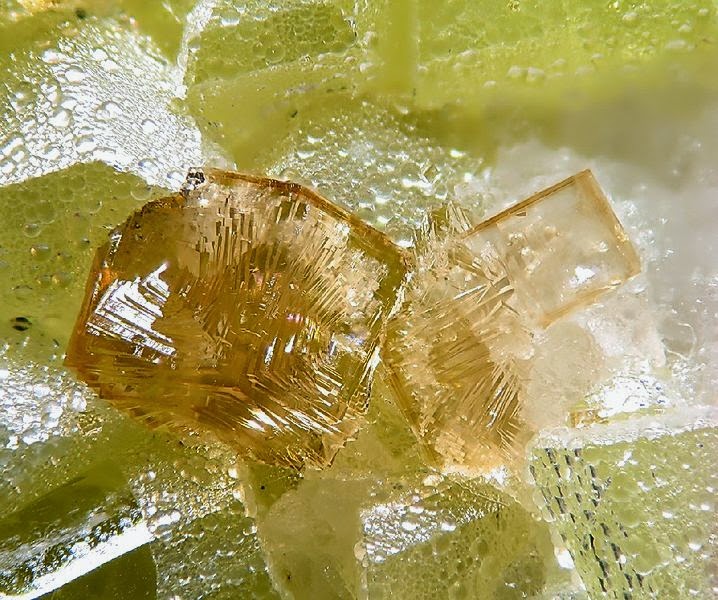
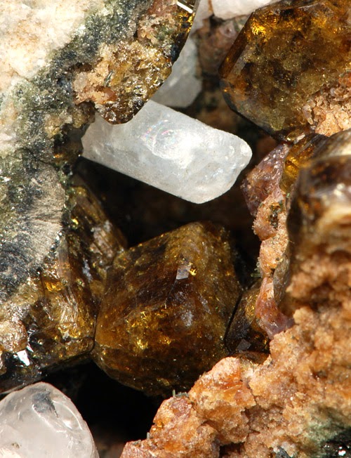
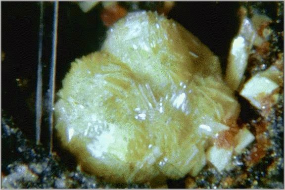
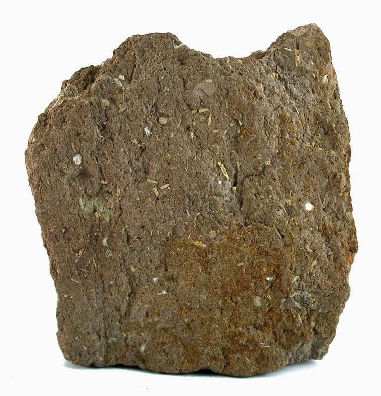
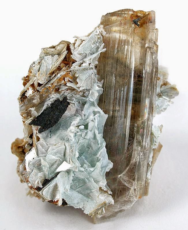
Chemical Formula: 46SiO2·6(N2,CO2)·2(CH4,N2) Locality: In Italy, at Solfatara Giona, Racalmuto, and at Caltanissetta, Sicily. Name Origin: From the Greek for “black” and “to be burned” in allusion to the fact that some specimens blacken on heating. Low temperature form.
Melanophlogite (MEP) is a rare silicate mineral and a polymorph of silica (SiO2). It has a zeolite-like porous structure which results in relatively low and not well-defined values of its density and refractive index. Melanophlogite often overgrows crystals of sulfur or calcite and typically contains a few percent of organic and sulfur compounds. Darkening of organics in melanophlogite upon heating is a possible origin of its name, which comes from the Greek for “black” and “to be burned”.
Occurrence
Melanophlogite is a rare mineral which usually forms round drops (see infobox) or complex intertwinned overgrowth structures over sulfur or calcite crystals. Rarely, it occurs as individual cubic crystallites a few millimeters in size. It is found in Parma, Torino, Caltanissetta and Livorno provinces of Italy; also in several mines of California in the US, in Crimea (Ukraine) and Pardubice Region (Czech Republic).
History
Discovery date : 1876 Town of Origin : SOLFATARE GIONA, RACALMUTO, SICILE Country of Origin : ITALIE
Optical properties
Optical and misc. Properties : Transparent to Translucent Refractive Index : from 1,42 to 1,45
Physical Properties
Cleavage: None Color: Brown, Colorless, Light yellow, Dark reddish brown. Density: 1.99 – 2.11, Average = 2.04 Diaphaneity: Transparent to Translucent Fracture: Brittle – Generally displayed by glasses and most non-metallic minerals. Hardness: 6.5-7 – Pyrite-Quartz Luminescence: Fluorescent, Short UV=weak gray-white, Long UV=gray-white. Luster: Vitreous (Glassy) Streak: white
Dean Wilson recently returned from a research cruise off Japan, carrying out deep-sea drilling to gather rock samples and sensor data on the geology beneath the seabed. The results will give us a better understanding of the risk of earthquakes and tsunamis. He describes life aboard the good ship Chikyu.
Ahead of my first trip to Japan, my head was full of childhood images of futuristic robots and high-speed trains. Tokyo didn’t disappoint. In the two days I had on dry land, I experienced delicious food, friendly people and the crazy juxtaposition of tranquil shrines in the midst of a busy city. It was a whirlwind experience.
The next morning, I found myself on a small passenger helicopter with a handful of other scientists heading out over the Philippine Sea, to a drop in the ocean about 100km south of Japan. Thirty minutes later I caught my first glimpse of the deep-sea drilling vessel Chikyu, essentially a mobile drilling platform.
It casts an unmistakable silhouette against the enormous expanse of the ocean. The growing image of the giant ship was stupendous. With its 70m derrick (drilling rig) standing proudly to attention in the centre of the vessel, it looked like a giant Tetris block sent down from the heavens! The Chikyu would be my home, office and lab for the next seven weeks. Suddenly a wave of emotions washed over me: I was excited, nervous and a little hysterical – what was I doing here?
About ten months earlier, I applied to sail on the Integrated Ocean Drilling Program’s (IODP) Expedition 338, a sea-going science mission to understand what causes large earthquakes and the generation of tsunami waves. Here’s what I thought when reading the advert: ‘WANTED: team of specialist scientists needed for intrepid exploration of the Earth below the sea. Seven weeks of hard but rewarding work out on the ocean waves. Beards optional!’
As a full-time researcher in marine geophysics, I spend most of my days sitting at a computer, so I really relish the opportunity to escape from the office and get some first-hand experience of collecting the data that is so crucial to my work.
Expedition 338 is part of a larger project aimed at learning more about how and why earthquakes and tsunamis occur. The IODP explores the geology below the seafloor to study Earth processes that evolve over time, ultimately causing violent, unpredictable natural disasters. The Nankai Trough Seismogenic Zone Experiment (NanTroSEIZE) is a complex ocean drilling project that is being conducted over several years (2007 to present) with multiple expeditions and scientists from all over the world.
NanTroSEIZE is the first attempt to drill, sample, and instrument the earthquake-causing or ‘seismogenic’ portion of the Earth’s crust, where violent, large-scale quakes have occurred repeatedly throughout history. The Nankai Trough is one of the most seismically active zones on the planet, and our sensors and sample data are expected to yield insights into the processes responsible for earthquakes and tsunamis, with implications for disaster planning and early warning systems.
Ice cream, ping-pong and borehole geophysics
Daily life onboard Chikyu was easy going. Meals are provided every six hours, washing is done within four and cabins are cleaned regularly. Everything is run to ensure that the ship’s crew, drilling engineers and scientists can work around the clock. The scientists have a daily meeting, with an operation and logistics update, science presentations, as well as morale-boosting items like choosing logo designs and planning the Christmas party – strictly no alcohol allowed though.
After several weeks, ‘Chikyu Time’ sets in, where days feel like weeks and every day is Groundhog Day. There are, however, plenty of things to break up the routine – ice cream twice a week, ping-pong tournaments, film screenings and even a sauna and hot tub.
Chikyu is an amazing machine. Using its six computer-controlled thrusters, the 210m, 57,000-tonne vessel can stay in exactly the same position for months at a time in all but the most challenging conditions. (For comparison, the Eiffel Tower weighs about 10,000 tonnes.) It can drill a staggering 7km below the seafloor, in water up to 2.5km deep. If the drill pipe that extends from the ship to the seafloor were as thick as a straw, it would be 100m long.
During Expedition 338, we drilled 12 holes into different parts of the seabed. They reached up to 2km below the seabed, and targeted different features identified from seafloor maps and images of the subsurface. At some holes we recovered rock samples (cores), while at others we measured geophysical properties, including electrical conductivity and acoustic velocity, from within the borehole while drilling. The holes were 30cm across – the size of a regular pizza – and we recovered the cores from inside the hollow drill barrel, known as the string, using a method akin to coring an apple.
In the end the recovered core is pulled up inside a core liner that’s about the same size as a household drainpipe. After this, the cores get split in two lengthways. One half is described and measured on board, with samples taken for later work, while the second half is archived. This involved categorising the sediments and rocks based on their mineralogy, elemental composition and grain size to understand where they came from – for example, from submarine river deposits or volcanic ash layers. Fossils and magnetic minerals can be used to understand the age of the material, and structures within it are analysed to understand how the rocks have been deformed since they were deposited.
My job was to interpret the geophysical data that were collected whilst drilling holes where no core samples were taken. This involved spending lots of time analysing curves and images for patterns and relating this information to what we already knew about the subsurface geology from the cores recovered at nearby holes. Once I’d analysed the data, key observations were compiled into reports that will eventually be used as an expedition reference volume for the whole scientific community.
Chikyu was also recently involved in IODP’s Japan Trench Fast Drilling Project (JFAST), to understand the very large fault slip that occurred in the shallow subseafloor during the 2011 Tohoku earthquake. (A fault slip is when two sections of the earth’s crust that were previously locked together by friction suddenly slide over each other.) This large slip of 30 to 50 metres was the main source of the devastating tsunami that caused so much damage and loss of life along the northeast coast of Honshu.
Understanding the Tohoku earthquake and tsunami has obvious benefits in evaluating the hazards at other subduction zones around the world. At these zones, the vast tectonic plates of the Earth’s crust are gradually sliding past each other, one beneath the other along the largest faults on Earth. Friction between the plates makes them grip together, building up energy, until they suddenly slip, releasing the stored energy in an earthquake. Obtaining a piece of the fault that moved tens of metres during the earthquake will provide meaningful new geological information. Scientists have never seen samples of a fault that has moved so far during a recent subduction zone earthquake.
Although Expedition 338 ended in January, there is still a great deal of work to be done. Our tasks include reports, meetings, post-cruise research, scientific publications and wider public outreach activities. Expeditions are expensive, but the rare data and samples we collected will be worked on for many years to come. When new techniques are developed or new theories need to be tested, the researchers of the future will be able to build on the work we did on the cruise to better understand the secrets of the Earth.
Note : The above story is based on materials provided by Dr Dean J Wilson is a marine geophysicist at the University of Southampton.
The paleoclimate record for the last ice age — a time 21,000 years ago called the “Last Glacial Maximum” (LGM) — tells of a cold Earth whose northern continents were covered by vast ice sheets. Chemical traces from plankton fossils in deep-sea sediments reveal rearranged ocean water masses, as well as extended sea ice coverage off Antarctica. Air bubbles in ice cores show that carbon dioxide in the atmosphere was far below levels seen before the Industrial Revolution.
While ice ages are set into motion by Earth’s slow wobbles in its transit around the sun, researchers agree that the solar-energy decrease alone wasn’t enough to cause this glacial state. Paleoclimatologists have been trying to explain the actual mechanism behind these changes for 200 years.
“We have all these scattered pieces of information about changes in the ocean, atmosphere, and ice cover,” says Raffaele Ferrari, the Breene M. Kerr Professor of Physical Oceanography in MIT’s Department of Earth, Atmospheric and Planetary Sciences, “and what we really want to see is how they all fit together.”
Researchers have always suspected that the answer must lie somewhere in the oceans. Powerful regulators of Earth’s climate, the oceans store vast amounts of organic carbon for thousands of years, keeping it from escaping into the atmosphere as CO2. Seawater also takes up CO2 from the atmosphere via photosynthesizing microbes at the surface, and via circulation patterns.
In a new application of ocean physics, Ferrari, along with Malte Jansen PhD ’12 of Princeton University and others at the California Institute of Technology, have found a new approach to the puzzle, which they detail in this week’s Proceedings of the National Academy of Sciences.
Lung of the ocean
The researchers focused on the Southern Ocean, which encircles Antarctica — a critical part of the carbon cycle because it provides a connection between the atmosphere and the deep ocean abyss. Ruffled by the winds whipping around Antarctica, the Southern Ocean is one of the only places where the deepest carbon-rich waters ever rise to the surface, to “breathe” CO2 in and out.
The modern-day Southern Ocean has a lot of room to breathe: Deeper, carbon-rich waters are constantly mixing into the waters above, a process enhanced by turbulence as water runs over jagged, deep-ocean ridges.
But during the LGM, permanent sea ice covered much more of the Southern Ocean’s surface. Ferrari and colleagues decided to explore how that extended sea ice would have affected the Southern Ocean’s ability to exchange CO2 with the atmosphere.
Shock to the system
This question demanded the use of the field’s accumulated knowledge of ocean physics. Using a mathematical equation that describes the wind-driven ocean circulation patterns around Antarctica, the researchers calculated the amount of water that was trapped under the sea ice by currents in the LGM. They found that the shock to the entire Earth from this added ice cover was massive: The ice covered the only spot where the deep ocean ever got to breathe. Since the sea ice capped these deep waters, the Southern Ocean’s CO2 was never exhaled to the atmosphere.
The researchers then saw a link between the sea ice change and the massive rearrangement of ocean waters that is evident in the paleoclimate record. Under the expanded sea ice, a greater amount of upwelled deep water sank back downward. Southern Ocean abyssal water eventually filled a greater volume of the entire midlevel and lower ocean — lifting the interface between upper and lower waters to a shallower depth, such that the deep, carbon-rich waters lost contact with the upper ocean. Breathing less, the ocean could store a lot more carbon.
A Southern Ocean suffocated by sea ice, the researchers say, helps explain the big drop in atmospheric CO2 during the LGM.
Dependent relationship
The study suggests a dynamic link between sea-ice expansion and the increase of ocean water insulated from the atmosphere, which the field has long treated as independent events. This insight takes on extra relevance in light of the fact that paleoclimatologists need to explain not just the very low levels of atmospheric CO2 during the last ice age, but also the fact that this happened during each of the last four glacial periods, as the paleoclimate record reveals.
Ferrari says that it never made sense to argue that independent changes drew down CO2 by the exact same amount in every ice age. “To me, that means that all the events that co-occurred must be incredibly tightly linked, without much freedom to drift beyond a narrow margin,” he says. “If there is a causality effect among the events at the start of an ice age, then they could happen in the same ratio.”
Note : The above story is based on materials provided by Massachusetts Institute of Technology.
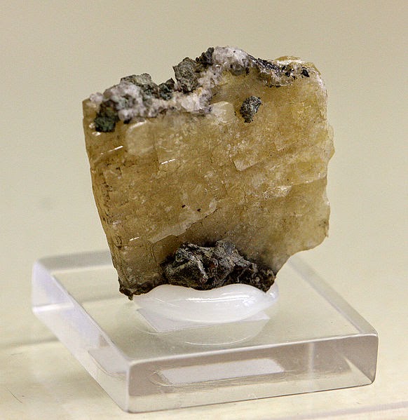
Chemical Formula: Ca4Al6Si6O24CO3 Locality: Mte. Somma, Vesuvius, Italy. Name Origin: From the Greek for “less”, referring to its less acute pyramidal form compared with vesuvianite.
Meionite is a tectosilicate belonging to the scapolite group with the formula Ca4Al6Si6O24CO3. Some samples may also contain a sulfate group. It was first discovered in 1801 on Mt Somma, Vesuvius, Italy.
History
Discovery date : 1801 Town of Origin : MONTE SOMMA, MT. VESUVE (VOLCAN), NAPLES, CAMPANIE Country of Origin: ITALIE
Optical properties
Optical and misc. Properties: Transparent to subtranslucent Refractive Index : from 1,55 to 1,60
Physical Properties
Cleavage: {???} Distinct, {???} Indistinct Color: Bluish, Brownish, Colorless, Violet, Greenish. Density: 2.66 – 2.73, Average = 2.69 Diaphaneity: Transparent to subtranslucent Fracture: Sub Conchoidal – Fractures developed in brittle materials characterized by semi-curving surfaces. Hardness: 5-6 – Between Apatite and Orthoclase Luminescence: Fluorescent, Short UV=yellow-white, Long UV=red. Luster: Vitreous – Resinous Streak: colorless
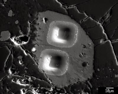
This shows secondary electron image of pits left by ion microprobe analyses of a heterogeneous apatite grain in Apollo sample 14321, 1047. Water has now been detected in apatite in many different lunar rock types. Credit: Katharine L. Robinson, University of Hawaii, HIGP
A recent review of hundreds of chemical analyses of Moon rocks indicates that the amount of water in the Moon’s interior varies regionally – revealing clues about how water originated and was redistributed in the Moon. These discoveries provide a new tool to unravel the processes involved in the formation of the Moon, how the lunar crust cooled, and its impact history.
This is not liquid water, but water trapped in volcanic glasses or chemically bound in mineral grains inside lunar rocks. Rocks originating from some areas in the lunar interior contain much more water than rocks from other places. The hydrogen isotopic composition of lunar water also varies from region to region, much more dramatically than in Earth.
The present consensus is that the Moon formed as the result of a giant impact of an approximately Mars-sized planetesimal with the proto-Earth. The water in the Moon is a tracer of the processes that operated in the hot, partly silicate gas, partly magma disk surrounding Earth after that impact.
The source of the Moon’s water has important implications for determining the source of Earth’s water, which is vital to life. There are two options: either, water was inherited by the Moon from the Earth during the Moon-forming impact, or it was added to the Moon later by comets or asteroids. It might also be a combination of these two processes.
“Basically, whatever happened to the Moon also happened to the Earth,” said Katharine Robinson, lead author of the study and Graduate Assistant at the University of Hawai’i – Mānoa (UHM) School of Ocean and Earth Science and Technology.
Robinson and Researcher G. Jeffrey Taylor, both at the UHM Hawai’i Institute of Geophysics and Planetology, compiled water measurements from lunar samples performed by colleagues from around the world, as well as their own. Specifically, they measured hydrogen and its isotope, deuterium (hydrogen with an extra neutron in its nucleus) with ion microprobes, which use a focused beam of ions to sputter ions from a small rock sample into a mass spectrometer. The ratio of hydrogen to deuterium can indicate the source of the water or trace magmatic processes in the lunar interior.
When water was first discovered in lunar samples in 2008, it was very surprising because from the time Apollo astronauts brought lunar samples, scientists thought that the Moon contained virtually no water.
“This was consistent with the idea that blossomed during the Origin of the Moon conference in Kona in 1984 — that the Moon formed by a giant impact with the still-growing Earth, leading to extensive loss of volatile chemicals. Our work is surprising because it shows that lunar formation and accretion were more complex than previously thought,” said Robinson.
The study of water in the Moon is still quite new, and many rocks have not yet been studied for water. The HIGP researchers have a new set of Apollo samples from NASA that they will be studying in the next few months, looking for additional clues about the early life of Earth and the Moon.
Note : The above story is based on materials provided by University of Hawaii ‑ SOEST
Bill Whittaker/Iowa Office of the State Archaeologist/Creative Commons
Things were looking up for Earth about 12,800 years ago. The last Ice Age was coming to an end, mammoths and other large mammals romped around North America, and humans were beginning to settle down and cultivate wild plants. Then, suddenly, the planet plunged into a deep freeze, returning to near-glacial temperatures for more than a millennium before getting warm again. The mammoths disappeared at about the same time, as did a major Native American culture that thrived on hunting them. A persistent band of researchers has blamed this apparent disaster on the impact of a comet or asteroid, but a new study concludes that the real explanation for the chill, at least, may lie strictly with Earth-bound events.
The study “pulls the rug out from under the contrived impact hypothesis quite nicely,” says Christian Koeberl, a geochemist at the University of Vienna. Most evidence for the extraterrestrial impact hypothesis, he says, was conjured up “out of thin air.”
The 1300-year big chill is known as the Younger Dryas, so called because of the sudden worldwide appearance of the cold-weather flowering plant Dryas octopetala. A number of causes have been suggested, including changes in ocean currents due to melting glaciers and volcanic activity. In 2007, a diverse group of 26 researchers, led by nuclear chemist Richard Firestone of the Lawrence Berkeley National Laboratory in California, formally proposed what is known as the Younger Dryas impact hypothesis, in which one or more extraterrestrial bodies blew up over North America, leading to widespread wildfires and strewing sun-blocking dust and debris across the globe.
In a series of papers, Firestone and his colleagues claimed various kinds of evidence for the hypothesis, including deposits of the element iridium (rare on Earth but abundant in meteorites), microscopic diamonds (called nanodiamonds), and magnetic particles in deposits at sites supposedly dated to about 12,800 years ago. The notion was popularized in television documentaries and other coverage on the National Geographic Channel, History Channel, and the PBS program NOVA. These claims were sharply contested by some specialists in the relevant fields, however, who either did not detect such evidence or argued that the deposits had other causes than a cosmic impact. For example, some say that nanodiamonds are common in ordinary geological formations, and that magnetic particles could come from ordinary fires.
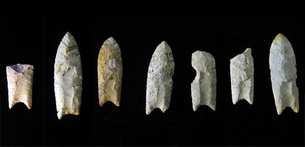
Now comes what some researchers consider the strongest attack yet on the Younger Dryas impact hypothesis. In a paper published online this week in the Proceedings of the National Academy of Sciences, a team led by David Meltzer, an archaeologist at Southern Methodist University, Dallas, in Texas, looks at the dating of 29 different sites in the Americas, Europe, and the Middle East in which impact advocates have reported evidence for a cosmic collision. They include sites in which sophisticated stone projectiles called Clovis points, used by some of the earliest Americans to hunt mammals beginning about 13,000 years ago, have been found, such as Chobot in Alberta, Canada, Murray Springs in Arizona, and Paw Paw Cove in Maryland; the site of Abu Hureyra in Syria, where evidence of plant-cultivating hunter-gatherers occurs; and sites in Greenland, Germany, Belgium, and the Netherlands where other evidence for an impact has been claimed. The team argues that when the quality and accuracy of the dating—which was based on radiocarbon and other techniques—is examined closely, only three of the 29 sites actually fall within the time frame of the Younger Dryas onset, about 12,800 years ago; the rest were probably either earlier or later by hundreds (and in one case, thousands) of years.
“The supposed Younger Dryas impact fails on both theoretical and empirical grounds,” says Meltzer, who adds that the popular appeal of the hypothesis is probably due to the way that it provides “simple explanations for complex problems.” Thus, “giant chunks of space debris clobbering the planet and wiping out life on Earth has undeniably broad appeal,” Meltzer says, whereas “no one in Hollywood makes movies” about more nuanced explanations, such as Clovis points disappearing because early Americans turned to other forms of stone tool technology as the large mammals they were hunting went extinct as a result of the changing climate or hunting pressure.
Maarten Blaauw, a paleoecologist at Queen’s University Belfast in the United Kingdom, finds the new work convincing. “It is vital to get the ages right,” he says, which “appears to have been lacking in the case of the [impact] papers” that Meltzer and his colleagues reanalyzed. “This paper should be read widely, and its lessons learned by the paleo community and by archaeologists.”
But impact proponents appear unmoved by the new study. “We still stand fully behind the [impact hypothesis], which is based on more than a confluence of dates,” Firestone says. “Radiocarbon dating is a perilous process,” he contends, adding that the presence of Clovis artifacts and mammoth bones just under the claimed iridium, nanodiamond, and magnetic sphere deposits is a more reliable indicator that an extraterrestrial event was responsible for their disappearance.
Note : The above story is based on materials provided by Michael Balter forAmerican Association for the Advancement of Science.
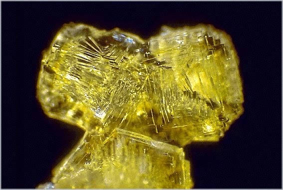
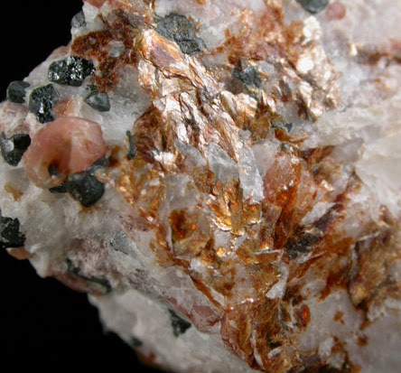
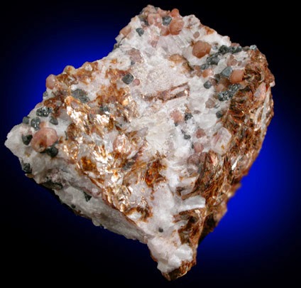
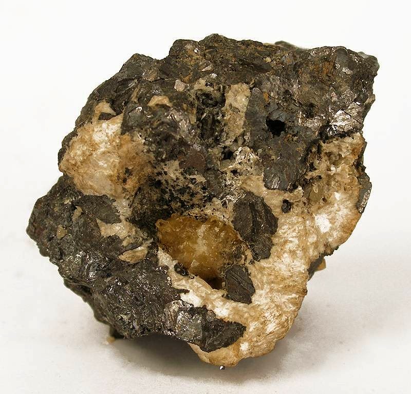
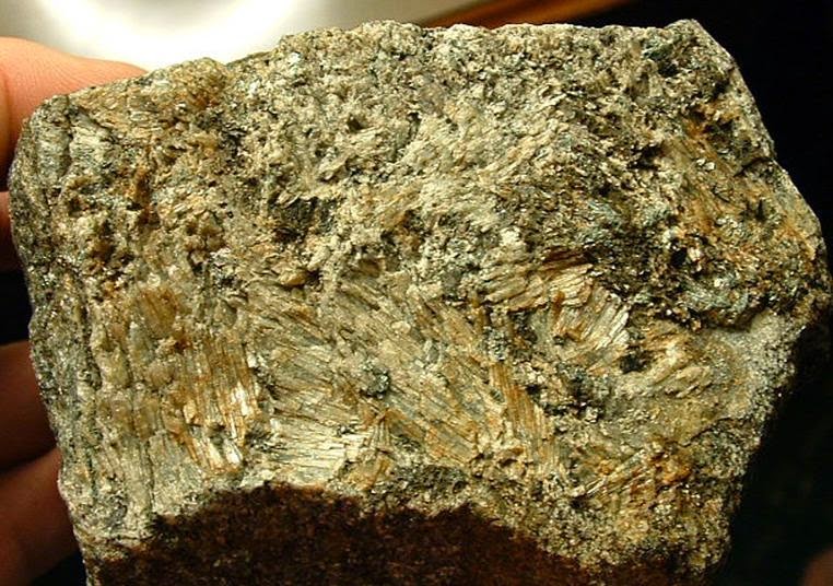
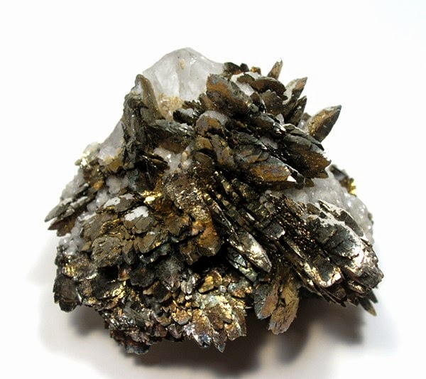
Chemical Formula: Mn19Zn3(AsO4)3(AsO3)(SiO4)3(OH)21 Locality: Sterling Hill, Franklin, Sussex Co., New Jersey, USA. Name Origin: Named for J. J. McGovern (1915-), Franklin miner and mineral collector.
History
Discovery date : 1927 Town of Origin : MINE STERLING HILL, OGDENSBURG, SUSSEX CO., NEW JERSEY Country of Origin : USA
Optical properties
Optical and misc. Properties : Translucent Refractive Index : from 1,75 to 1,76
Physical Properties
Cleavage: {001} Perfect Color: Bronze brown, Light brown, Dark reddish brown, Reddish brown. Density: 3.72 Diaphaneity: Translucent Fracture: Brittle – Generally displayed by glasses and most non-metallic minerals. Luminescence: Non-fluorescent. Luster: Vitreous – Pearly Streak: brown
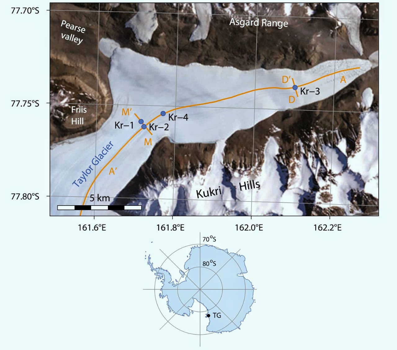
Top: satellite imagery of Taylor Glacier. Kr-81 sampling locations are indicated as blue dots. Bottom: location of Taylor Glacier on map of Antarctica. Image credit: Christo Buizert et al.
The new technique is much like the more-heralded carbon-14 dating technique that measures the decay of a radioactive isotope and compares it to a stable isotope.
Unlike carbon-14, however, krypton does not interact chemically and is much more stable with a half-life of around 230,000 years.
Carbon dating doesn’t work well on ice because carbon-14 is produced in the ice itself by cosmic rays and only goes back some 50,000 years.
Krypton is produced by cosmic rays bombarding our planet and then stored in air bubbles trapped within ice. It has a radioactive isotope, krypton-81, that decays very slowly, and a stable isotope (krypton-83) that does not decay.
Comparing the proportion of stable-to-radioactive isotopes provides the age of the ice.
In their study, reported in the Proceedings of the National Academy of Sciences, Dr Buizert with colleagues put four 350-kg samples of ice into a container and melted it to release the air from the bubbles. The krypton was isolated from the air and sent for krypton-81 counting.
They determined from the isotope ratio that the Taylor Glacier samples were 120,000 years old, and validated the estimate by comparing the results to well-dated ice core measurements of atmospheric methane and oxygen from that same period.
Now the challenge is to locate some of the oldest ice in Antarctica, which may not be as easy as it sounds.
“Most people assume that it’s a question of just drilling deeper for ice cores, but it’s not that simple. Very old ice probably exists in small isolated patches at the base of the ice sheet that have not yet been identified, but in many places it has probably melted and flowed out into the ocean,” explained co-author Dr Edward Brook of Oregon State University.
The scientists are hoping that the new technique will help identify ice that is more than a million years old, thereby reconstructing climate much farther back into Earth’s history.
“Reconstructing the Earth’s climate back to 1.5 million years is important because a shift in the frequency of ice ages took place in what is known as the Middle Pleistocene transition. The Earth is thought to have shifted in and out of ice ages every 100,000 years or so during the past 800,000 years, but there is evidence that such a shift took place every 40,000 years prior to that time,” Dr Buizert said.
“Why was there a transition from a 40,000-year cycle to a 100,000-year cycle? Some people believe a change in the level of atmospheric carbon dioxide may have played a role. That is one reason we are so anxious to find ice that will take us back further in time so we can further extend data on past carbon dioxide levels and test this hypothesis,” he concluded.
More Information : Christo Buizert et al. Radiometric 81Kr dating identifies 120,000-year-old ice at Taylor Glacier, Antarctica. PNAS, published online April 21, 2014; doi: 10.1073/pnas.1320329111
Note : The above story is based on materials provided by Sci-News
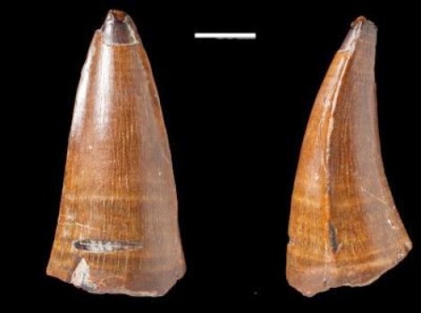
This is a tooth recovered from Chesil Beach in Dorset, England, which belonged to a Dakosaurus. Credit: Mark Young and Lorna Steel
A fossilised tooth belonging to a fearsome marine predator has been recorded as the largest of its kind found in the UK, following its recent discovery.
A team of palaeontologists have verified the tooth, which was found near Chesil Beach in Dorset, as belonging to a prehistoric relative of modern crocodiles known as Dakosaurus maximus.
The tooth, which has a broken tip, is approximately 5.5 cm long.
Researchers and curators from University of Edinburgh and the Natural History Museum in London identified the item after it was bought at an online auction by a fossil collector.
Scientists say the circumstances in which the fossil was found were unusual — it was dredged from the sea floor rather than being found on the shore or dug up.
The tooth has been examined and identified by a team of UK palaeontologists and placed in the fossil collection of the Natural History Museum.
Dakosaurus maximus, which grew up to about 4.5 metres long, swam in the shallow seas that covered Europe some 152 million years ago. It belonged to a family of marine animals known as thalattosuchians, relatives of today’s crocodiles.
The unusual shape of the animal’s skull and teeth suggests it ate similar prey to modern-day killer whales. It would have used its broad, short jaws to swallow large fish whole and to bite chunks from larger prey.
The team’s research is published in the scientific journal Historical Biology.
Dr Mark Young, of the University of Edinburgh’s School of Biological Sciences, said: “Given its size, Dakosaurus had very large teeth. However, it wasn’t the top marine predator of its time, and would have swum alongside other larger marine reptiles, making the shallow seas of the Late Jurassic period exceptionally dangerous.”
Note : The above story is based on materials provided by University of Edinburgh.
Chemical Formula: PbFCl Locality: Cromford, near Matlock, Derbyshire. Ancient lead slags at Laurium, Greece. Name Origin: Named from its locality.
Matlockite is a rare lead halide mineral, named after the town of Matlock in Derbyshire, England, where it was first discovered in a nearby mine. Matlockite (chemical formula: PbFCl) gives its name to the matlockite group which consists of rare minerals of a similar structure.
Description
The mineral, a lead fluorochloride (formula PbFCl), was discovered sometime around the early 1800s at Bage Mine at Bolehill near Matlock, together with specimens of phosgenite and anglesite. Although phosgenite was known at this time, it seems likely that matlockite itself remained unappreciated as a new mineral for some fifty years. It was given the name by Greg in 1851. The first mention of Matlockite may have been in Mawe’s Mineralogy of Derbyshire in 1802 in which he gives a detailed description of phosgenite, which is then followed by a mention of a mineral he refers to as “glass lead” – a description which does rather equate to the appearance of matlockite. It is a light, translucent creamy-yellow colour, but heavy in weight having a density that is over 7.1.
A very large specimen 10 cm across, and originating from Derbyshire, exists in the collections of the American Museum of Natural History. A 7 cm specimen can be found in the collection of Derby Museum and Art Gallery.
Matlockite has been reported from a variety of locations since its discovery at the type locality of Derbyshire. The mineral is also found in Tiger, Arizona, Laurium in Greece, a mine near Essen in Germany and near Campiglia in Tuscany. Samples have also been found at locations in South Africa, Peru, Chile, Australia, Austria, France and Italy.
Optical properties
Optical and misc. Properties : Transparent Refractive Index : from 2,00 to 2,14
Physical Properties
Cleavage: {001} Perfect Color: Brownish yellow, Colorless, Green yellow, Dark yellow brown. Density: 7.12 Diaphaneity: Transparent Fracture: Brittle – Uneven – Very brittle fracture producing uneven fragments. Hardness: 2.5-3 – Finger Nail-Calcite Luminescence: Non-Fluorescent. Luster: Adamantine – Pearly Streak: white
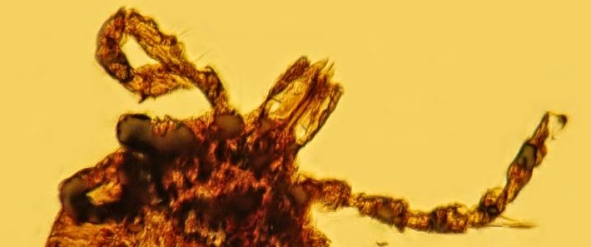
Tick carrying spirochetes. Credit: Image courtesy of Oregon State University
Lyme disease is a stealthy, often misdiagnosed disease that was only recognized about 40 years ago, but new discoveries of ticks fossilized in amber show that the bacteria which cause it may have been lurking around for 15 million years — long before any humans walked on Earth.
The findings were made by researchers from Oregon State University, who studied 15-20 million-year-old amber from the Dominican Republic that offer the oldest fossil evidence ever found of Borrelia, a type of spirochete-like bacteria that to this day causes Lyme disease. They were published in the journal Historical Biology.
In a related study, published in Cretaceous Research, OSU scientists announced the first fossil record of Rickettsial-like cells, a bacteria that can cause various types of spotted fever. Those fossils from Myanmar were found in ticks about 100 million years old.
As summer arrives and millions of people head for the outdoors, it’s worth considering that these tick-borne diseases may be far more common than has been historically appreciated, and they’ve been around for a long, long time.
“Ticks and the bacteria they carry are very opportunistic,” said George Poinar, Jr., a professor emeritus in the Department of Integrative Biology of the OSU College of Science, and one of the world’s leading experts on plant and animal life forms found preserved in amber. “They are very efficient at maintaining populations of microbes in their tissues, and can infect mammals, birds, reptiles and other animals.
“In the United States, Europe and Asia, ticks are a more important insect vector of disease than mosquitos,” Poinar said. “They can carry bacteria that cause a wide range of diseases, affect many different animal species, and often are not even understood or recognized by doctors.
“It’s likely that many ailments in human history for which doctors had no explanation have been caused by tick-borne disease.”
Lyme disease is a perfect example. It can cause problems with joints, the heart and central nervous system, but researchers didn’t even know it existed until 1975. If recognized early and treated with antibiotics, it can be cured. But it’s often mistaken for other health conditions. And surging deer populations in many areas are causing a rapid increase in Lyme disease — the confirmed and probable cases of Lyme disease in Nova Scotia nearly tripled in 2013 over the previous year.
The new research shows these problems with tick-borne disease have been around for millions of years.
Bacteria are an ancient group that date back about 3.6 billion years, almost as old as the planet itself. As soft-bodied organisms they are rarely preserved in the fossil record, but an exception is amber, which begins as a free-flowing tree sap that traps and preserves material in exquisite detail as it slowly turns into a semi-precious mineral.
A series of four ticks from Dominican amber were analyzed in this study, revealing a large population of spirochete-like cells that most closely resemble those of the present-day Borrelia species. In a separate report, Poinar found cells that resemble Rickettsia bacteria, the cause of Rocky Mountain spotted fever and related illnesses. This is the oldest fossil evidence of ticks associated with such bacteria.
In 30 years of studying diseases revealed in the fossil record, Poinar has documented the ancient presence of such diseases as malaria, leishmania, and others. Evidence suggests that dinosaurs could have been infected with Rickettsial pathogens.
Humans have probably been getting diseases, including Lyme disease, from tick-borne bacteria as long as there have been humans, Poinar said. The oldest documented case is the Tyrolean iceman, a 5,300-year-old mummy found in a glacier in the Italian Alps.
“Before he was frozen in the glacier, the iceman was probably already in misery from Lyme disease,” Poinar said. “He had a lot of health problems and was really a mess.”
Note : The above story is based on materials provided by Oregon State University.
A Curtin University researcher has shown that ancient volcanic eruptions in Australia 510 million years ago significantly affected the climate, causing the first known mass extinction in the history of complex life.
Published in the journal Geology, Associate Professor Fred Jourdan from Curtin’s Department of Applied Geology, along with colleagues from several Australian and international institutions, used radioactive dating techniques to precisely measure the age of the eruptions of the Kalkarindji volcanic province — where lavas covered an area of more than 2 million square kilometres in the Northern Territory and Western Australia.
Dr Jourdan and his team were able to prove the volcanic province occurred at the same time as the Early-Middle Cambrian extinction from 510-511 million years ago — the first extinction to wipe out complex multicellular life.
“It has been well-documented that this extinction, which eradicated 50 per cent of species, was related to climatic changes and depletion of oxygen in the oceans, but the exact mechanism causing these changes was not known, until now,” Dr Jourdan said.
“Not only were we able to demonstrate that the Kalkarindji volcanic province was emplaced at the exact same time as the Cambrian extinction, but were also able to measure a depletion of sulphur dioxide from the province’s volcanic rocks — which indicates sulphur was released into the atmosphere during the eruptions.
“As a modern comparison, when the small volcano Pinatubo erupted in 1991, the resulting discharge of sulphur dioxide decreased the average global temperatures by a few tenths of a degree for a few years following the eruption.
“If relatively small eruptions like Pinatubo can affect the climate just imagine what a volcanic province with an area equivalent to the size of the state of Western Australia can do.”
The team then compared the Kalkarindji volcanic province with other volcanic provinces and showed the most likely process for all the mass extinctions was a rapid oscillation of the climate triggered by volcanic eruptions emitting sulphur dioxide, along with greenhouse gases methane and carbon dioxide.
“We calculated a near perfect chronological correlation between large volcanic province eruptions, climate shifts and mass extinctions over the history of life during the last 550 million years, with only one chance over 20 billion that this correlation is just a coincidence,” Dr Jourdan said.
Dr Jourdan said the rapid oscillations of the climate produced by volcanic eruptions made it difficult for various species to adapt, ultimately resulting in their demise. He also stressed the importance of this research to better understand our current environment.
“To comprehend the long-term climatic and biological effects of the massive injections of gas in the atmosphere by modern society, we need to recognise how climate, oceans and ecosytems were affected in the past,” he said.
Note : The above story is based on materials provided by Curtin University.
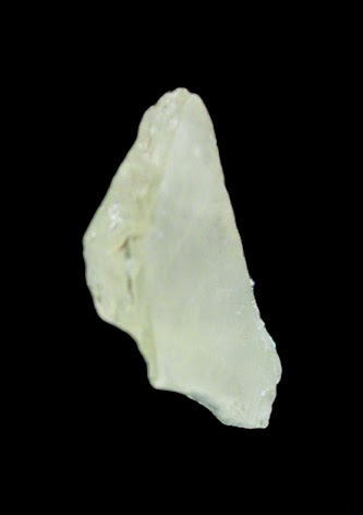
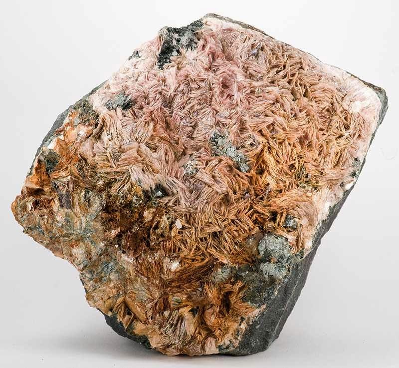
Chemical Formula: Na4Al3Si9O24Cl Locality: Pianura. Near Naples, Italy. Name Origin: Named by von Rath in honor of his wife, Maria Rosa vom Rath (1830-1888).
Marialite is a silicate mineral with a chemical composition of Na4Al3Si9O24Cl if a pure endmember or Na4(AlSi3O8)3(Cl2,CO3,SO4) with increasing meionite content. Marialite is a member of the scapolite group and a solid solution exists between marialite and meionite, the calcium endmember. It is a rare mineral usually used as a collector’s stone. It has a very rare but attractive gemstones and cat’s eye.
Discovery and occurrence
Marialite was first described in 1866 for an occurrence in the Phlegrean Volcanic complex, Campania, Italy. It was named by German mineralogist Gerhard vom Rath for his wife, Maria Rosa vom Rath.
Marialite occurs in regional and contact metamorphism: marble, calcareous gneiss, granulite and greenschist. It also occurs in skarn, pegmatite and hydrothermally altered volcanic rocks. This means that Marialite is formed in high pressure and/or high temperature environments.
History
Discovery date : 1866 Town of Origin : PIANURA, NAPLES, CAMPANIE Country of Origin : ITALIE
Optical properties
Optical and misc. Properties : Transparent to Translucent Refractive Index : from 1,53 to 1,55
Physical Properties
Cleavage: {100} Distinct, {110} Distinct Color: Bluish, Brownish, Colorless, Violet, Greenish. Density: 2.5 – 2.62, Average = 2.56 Diaphaneity: Transparent to Translucent Fracture: Brittle – Conchoidal – Very brittle fracture producing small, conchoidal fragments. Hardness: 5.5-6 – Knife Blade-Orthoclase Luminescence: Fluorescent, Long UV=strong yellow. Luster: Vitreous – Pearly Streak: white
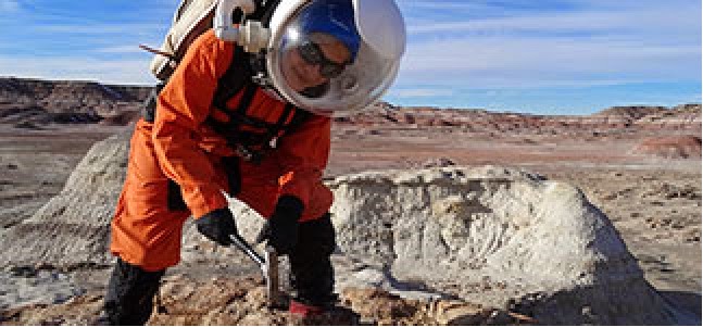
Some scientists may dream of the chance to pursue their research on another planet. That opportunity isn’t a reality just yet, but PhD student Michaela Musilova got the next best thing – a simulated mission to Mars.
Space suit fitted: check. Helmet secured: check. Radio transmitter attached: check. Air supply pack turned on: check. Time to go into the airlock! While the simulated depressurisation of the airlock is ending, my fellow crewmember and I finish making our plans for the EVA – extra vehicular activity.
I look through the porthole eagerly, in anticipation of stepping out onto the Martian terrain. It’s another sunny day on Mars, even though the temperature is still below zero. It’s a good thing our suits are thick enough to protect us against the cold, but that makes them very heavy: along with the air-supply pack they weigh 15kg.
As I walk over the rolling, red sandy hills of the stunning Martian landscape, I look back at the Mars Desert Research Station (MDRS). It is an analogue (simulation) laboratory – a copy of a planned NASA surface base on Mars – built by the non-profit Mars Society, which works closely with NASA and other international space agencies.
The station is in the high, cold Utah desert, USA, where the environmental conditions, geological features and biological attributes are a good approximation of what we know about those on Mars. It was designed to help people learn about the challenges of living and working on Mars. The Red Planet is considered to be the nearest planet with the resources for humanity to inhabit and then to use as a stepping stone for expansion further into the universe.
I am one of several scientists selected to take part in a total immersion simulation for over two weeks. This means we spend every minute of every day facing the physical and social challenges of life as we would experience it on Mars.
We are here as analogue astronauts, subjected to psychological, nutritional and scientific studies designed by researchers from around the world. These include living with limited amounts of electricity, oxygen, water and dehydrated powder-like astronaut’s food. Crews for simulations are selected to include specialists in different fields of research that would be necessary for the exploration of Mars.
Our crew commander is an aerospace engineer; we have a medical officer, two crew engineers, each specialising in a different aspect of space technology equipment testing; two crew scientists: a geochemist and myself – an astrobiologist and geologist. We also have a film-maker and even a humanoid robot, which we are testing out as a potential crew-member for real Mars missions.
We arrived at MDRS on 18 January 2014 and spent every moment of the simulation in total isolation from our terrestrial lives. The facility became our new home. It is made up of a habitat module nick-named the Hab, a greenhouse called the GreenHab and an observatory. The Hab is a two-storey cylinder-shaped building made to fit atop a heavy-lift space-launch vehicle. It’s only eight metres in diameter, creating a very confined living and working environment.
The common room, on the top floor, also serves as dining room, workstation, kitchen and exercise area. The lower level contains the airlock, laboratories, bathroom and toilet, all crammed into a space the size of my living room. As a consequence, you are almost always within eyeshot and earshot of at least one other person, so, it was really important that the crew could work as a team and get along for a prolonged period despite the lack of privacy.
Each of us has our own scientific experiments to conduct, which involve lab and/or field work in the simulated Martian environment. They include field-testing NASA hardware that extracts hydrogen and oxygen from soil, a technology that could potentially produce breathable air, drinkable water and rocket fuel for a return journey to Earth. If it works, this equipment would dramatically reduce the weight and cost of future space missions.
Our team also carried out simulated surgeries, via Skype, with several research groups around the world including the French/Italian Concordia base in Antarctica. The goal of these ‘tele-surgeries’ was to understand the difficulties faced when medical experts have to direct non-experts in an emergency, with restricted and delayed telecommunications – a situation astronauts travelling to Mars are likely to find themselves in.
One of the engineering projects is on prototype spacesuit glove technology. Our gloves have to be thick but these prototypes are nevertheless designed to feed information to the user’s fingertips about the texture and temperature of what they are holding, giving the astronaut a better awareness of the samples they are handling and the environment around them.
Another engineering project is testing rover cameras and a 3D mapping system similar to the one that will be on the ExoMars rover (scheduled for launch to Mars in 2018).
My own research is on extremophiles: organisms that live in physically or chemically extreme environments and are therefore significant for understanding what kind of life might exist on other planets and to help us develop the technologies to search for it. At MDRS, I am investigating two important questions: whether terrestrial microbes can survive in Mars-like conditions and thus whether there could be similar life on Mars; and whether these extremophiles could be used for terraforming Mars – recreating Earth-like conditions. Without terraforming or some other way of creating conditions for agriculture on Mars we could never properly settle on the Red Planet.
Today, I set out to collect more extremophiles for my experiments in the Hab (and for further analysis during my PhD). I love doing this simulated Martian fieldwork. Regardless of how hard it is to move around in my heavy spacesuit, breathing artificial air in my fishbowl helmet, I am completely absorbed in my role as an analogue astronaut. Walking towards the red Martian hills on the horizon I feel, more than ever, that I am on the path towards my childhood dream of going into space for real.
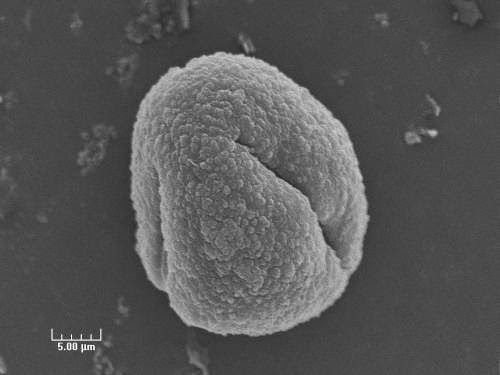
Single oak pollen grain, SEM image. Credit: Allison Steiner (UM) and Michael Pendleton (Texas A&M)
In the past, many atmospheric scientists believed that pollen particles probably had a negligible effect on climate because they were so big. In recent years, however, as they began to realize that pollen particles were not as sturdy as they once thought, they have been rethinking their old assumptions.
“Pollen can rupture and generate a lot of small, tiny particles,” says Allison Steiner, an associate professor of atmospheric, oceanic and space sciences at the University of Michigan. “They can break pretty easily.”
Moreover, pollen, the same airborne material that wreaks misery during certain seasons in the form of drippy noses and itchy eyes, apparently can have an influence on weather. When big pollen particles break into fine ones, they can take up water vapor in the air to promote the formation of clouds, potentially altering weather systems as a result. Unlike greenhouse gases, which contribute to warming, these fine particles can have a cooling effect.
This is a process that Steiner wants to learn more about, particularly now, when much of the scientific community is devoting considerable attention to the anthropogenic—or human—causes of climate change.
“The impact of pollen in the atmosphere may change weather and it could change our understanding of the climate system,” says the National Science Foundation (NSF)-funded scientist.
“How much is nature contributing?” she adds. “How important will that be in understanding what we will see in the absence of human influences? It’s easier to understand the human causes, but these natural aerosols like pollen are something we don’t understand very well.”
Prior research indicates that when pollen becomes wet, it easily ruptures into very small particles. She wondered whether these small, pollen fragments could, “seed” the creation of clouds.
“If you have water vapor in the atmosphere, it’s hard to form droplets all by itself,” she explains. “But if you have a little particle already there, it’s easy for water to condense on it and grow into a droplet, which enables the formation of cloud droplets.
“Most people think of pollen as being pretty inert in the atmosphere, and it’s not,” she adds. “It’s interacting with the water cycle, and can influence clouds in ways that people hadn’t realized before.”
She and her team are using ground based observation data obtained from across the nation to design a computer algorithm emissions model. The model includes the different types of pollen, and takes into account various conditions that can have an effect on pollen when it enters the atmosphere, for example, rain.
Furthermore, tiny pollen particles can react with radiation. “The models simulate the ability of pollen particles to interact with incoming solar radiation to understand how these particles will affect climate,” she says. By using computer models, she can estimate the effect these particles have on regional climate.
She also has been working in the laboratory of Sarah Brooks, a professor of atmospheric sciences at Texas A&M University, to demonstrate pollen’s effect on cloud formation. Using a cloud condensation nuclei chamber, an instrument that can reproduce the atmospheric conditions that form clouds, they were able to demonstrate that pollen can in fact grow and act as cloud droplets.
“This means that pollen could have an impact on climate,” says Steiner, who conducted the experiments at Texas A & M in the spring. “One thing we are still trying to figure out is how big that effect actually is.”
Steiner is conducting her research under an NSF Faculty Early Career Development (CAREER) award, which she received in 2010. The award supports junior faculty who exemplify the role of teacher-scholars through outstanding research, excellent education, and the integration of education and research within the context of the mission of their organization. NSF is funding her work with $599,940 over five years.
As part of the grant’s educational component, she has worked with middle schools and high schools in Detroit and Ypsilanti. Using the sites and numerous hands-on activities will introduce students to hypothesis development, data collection and analysis, and interpretation, and also will help the pollen emissions model development.
She also plans to integrate elements of the pollen project with University of Michigan undergraduate and graduate programs, as well as form a partnership with the International Centre for Theoretical Physics in Trieste, Italy to train scientists from developing nations on the role of biosphere-atmosphere interactions.
Steiner says she is especially gratified by the response of the young middle school students “who find it a real change to have a college professor come into their classroom on a regular basis,” she says, adding: “It can be a real challenge to make our research relevant for middle-school students. But the students have asked great questions, and we’ve developed some novel hands-on activities that have really helped the students to see how fun and exciting scientific research can be.”
Note : The above story is based on materials provided by National Science Foundation
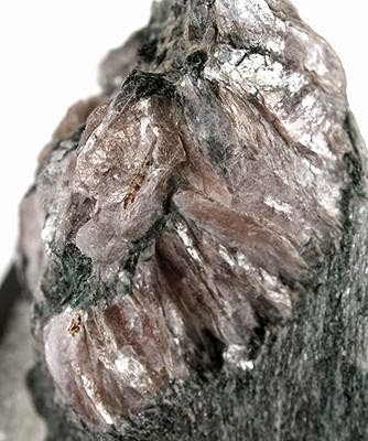
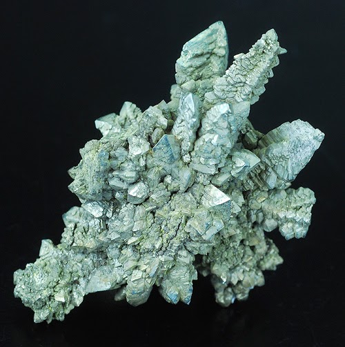
Chemical Formula: CaAl2(Al2Si2O10)(OH)2 Locality: Corundum mines at Ekaterinurg Distict, Ural Mountains, Russia. Name Origin: From the Greek margaritos – “pearl.”
Margarite is a calcium rich member of the mica group of the phyllosilicates with formula: CaAl2(Al2Si2O10)(OH)2. It forms white to pinkish or yellowish gray masses or thin laminae. It crystallizes in the monoclinic crystal system. It typically has a specific gravity of around 3 and a Mohs hardness of 4. It is translucent with perfect 010 cleavage and exhibits crystal twinning.
It occurs commonly as an alteration product of corundum, andalusite and other aluminous minerals. It has been reported as forming alteration pseudomorphs of chiastolite along with muscovite and paragonite. The margarite in this occurrence forms preferentially along the dark graphite rich inclusions with the chiastolite crystals.
History
Discovery date : 1823 Town of Origin: MT. GREINER, STERZING, TYROL Country of Origin : AUTRICHE
Optical properties
Optical and misc. Properties : Translucent to subtranslucent Refractive Index: from 1,63 to 1,65 Axial angle 2V : 40-67°
Physical Properties
Cleavage: {001} Good Color: White, Gray, Pinkish gray, Yellowish gray. Density: 2.99 – 3.08, Average = 3.03 Diaphaneity: Translucent to subtranslucent Fracture: Brittle – Generally displayed by glasses and most non-metallic minerals. Hardness: 4 – Fluorite Luminescence: Fluorescent, Short UV=sky blue, Long UV=strong sky blue. Luster: Pearly Streak: white
Volcanic eruptions across a vast area of what is now Western Australian and the Northern Territory 510 million years ago caused the first known mass extinction of complex life forms.
Curtin University’s Dr Fred Jourdan says it is widely documented that the Early-Middle Cambrian extinction of complex multicellular life was related to changes in climate and depletion of oxygen in the oceans but the exact cause has been unknown until now.
He is part of an international team of scientists that calculated a near perfect chronological correlation between large volcanic eruptions, climate changes and mass extinction over the history of life during the last 550 million years.
The eruptions produced rapid fluctuations in climate making it difficult for species to survive.
The research team’s findings High-precision dating of the Kalkarindji large igneous province, Australia, and synchrony with the early–Middle Cambrian (Stage 4–5) extinction, have been reported in the journal Geology.
The paper concludes the likely factors responsible for the Early–Middle Cambrian extinction are rapid climate shifts triggered by volcanic eruptions emitting mantle gases sulphur dioxide and greenhouse gases methane and carbon dioxide, either dissolved in the magma or generated by the interaction between magma and evaporite layers and/or oil-rich rocks.
Primative ocean life
Since there was no existing fauna or flora on land during the period, the extinction mechanism must have acted on the oceans.
Life at that time would have included the reef building sponge-like organism Archaeocyathids and Trilobites, the most primitive groups.
Dating techniques
The team used high-precision 40Ar/39Ar and U-Pb mineral dating to measure the age of eruptions in the Kalkarindji volcanic province in the Northern Territory and Western Australia where lavas covered more than two million square kilometres.
Both techniques are based on completely different elements and give the same age for the lavas, which is a strong validation that the age is correct.
Insights into gas emission effect
Dr Jourdan says the research is vital to understanding the long term implications that modern-day massive gas emission into the atmosphere can have on the climate and life.
“I’m talking about greenhouses gases like methane and carbon dioxide which warm the climate and sulphur dioxide which can cool the climate for short periods of time but more relevant to now, can cause acid rains which can wreck ecosystem and massive toxic pollution; part of the irritant pollutants in Beijing come from sulphur dioxide.
“…we can see the effect of those gases on nature by studying the rock record, and we are injecting a massive amount of those into the atmosphere, mostly by burning fossil fuels like coal and oil.”
More information: F. Jourdan, K. Hodges, B. Sell, U. Schaltegger, M.T.D. Wingate, L.Z. Evins, U. Söderlund, P.W. Haines, D. Phillips, and T. Blenkinsop. “High-precision dating of the Kalkarindji large igneous province, Australia, and synchrony with the Early–Middle Cambrian (Stage 4–5) extinction.” Geology, G35434.1, first published on April 24, 2014, DOI: 10.1130/G35434.1
Note : The above story is based on materials provided by Science Network WA
Scientists have found a way to extend the length of time they can use protein molecules to identify tissues like bone and teeth.
Every animal has thousands of proteins in its bones containing information on its species. These proteins make up an almost unique fingerprint that survives long after the animal’s DNA has decomposed.
Scientists can take advantage of this long survival time and use the proteins to try to identify fossils and other ancient tissues like hair and skin. But over time these proteins also begin to break down, which makes it hard to extract the genetic information they hold.
Last year Dr Mike Buckley of the University of Manchester showed that collagen, which contains almost 90 per cent of the proteins in bone, could be used to identify extremely old fossils when his collagen-fingerprinting technique identified a 1.5 million-year-old camel fossil from the high Arctic.
But proteins that aren’t found in collagen have only ever been used to test the genetic information of fossils up to 50,000 years old.
Now Buckley and his PhD student Caroline Wadsworth have managed to use these non-collagenous proteins to test fossils over one million years old.
They found that thousands of non-collagenous proteins – with the potential to be even more informative than collagen – also survive the burial process, and hope to use these in the future to test many older fossils.
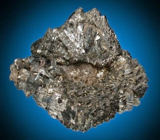
Chemical Formula: FeS2 Locality: Common world wide. Name Origin: Arabic or Moorish name for pyrites and similar material of uncertain origin.
The mineral marcasite, sometimes called white iron pyrite, is iron sulfide (FeS2) with orthorhombic crystal structure. It is physically and crystallographically distinct from pyrite, which is iron sulfide with cubic crystal structure. Both structures do have in common that they contain the disulfide S22- ion having a short bonding distance between the sulfur atoms.
The structures differ in how these di-anions are arranged around the Fe2+ cations. Marcasite is lighter and more brittle than pyrite. Specimens of marcasite often crumble and break up due to the unstable crystal structure.
On fresh surfaces it is pale yellow to almost white and has a bright metallic luster. It tarnishes to a yellowish or brownish color and gives a black streak. It is a brittle material that cannot be scratched with a knife. The thin, flat, tabular crystals, when joined in groups, are called “cockscombs.”
In marcasite jewellery, pyrite used as a gemstone is termed “marcasite”. That is, marcasite jewellery is made from pyrite not from marcasite. In the late medieval and early modern eras the word “marcasite” meant both pyrite and marcasite (and iron sulfides in general). The narrower, modern scientific definition for marcasite as orthorhombic iron sulfide dates from 1845. The jewellery sense for the word pre-dates this 1845 scientific redefinition. Marcasite in the scientific sense is not used as a gem due to its brittleness.
Occurrence
Marcasite can be formed as both a primary or a secondary mineral. It typically forms under low-temperature highly acidic conditions. It occurs in sedimentary rocks (shales, limestones and low grade coals) as well as in low temperature hydrothermal veins. Commonly associated minerals include pyrite, pyrrhotite, galena, sphalerite, fluorite, dolomite and calcite.
As a primary mineral it forms nodules, concretions and crystals in a variety of sedimentary rock, such as at Dover, Kent, England, where it forms as sharp individual crystals and crystal groups, and nodules (similar to those shown here) in chalk.
As a secondary mineral it forms by chemical alteration of a primary mineral such as pyrrhotite or chalcopyrite.
History
Discovery date : 1845
Optical properties
Optical and misc. Properties : Opaque Reflective Power : 49,1-54,1% (580)
Physical Properties
Cleavage: {010} Indistinct Color: Bronze, Light brass yellow, Tin white. Density: 4.89 Diaphaneity: Opaque Fracture: Uneven – Flat surfaces (not cleavage) fractured in an uneven pattern. Hardness: 6-6.5 – Orthoclase-Pyrite Luminescence: Non-fluorescent. Luster: Metallic Magnetism: Magnetic after heating Streak: gray brownish black
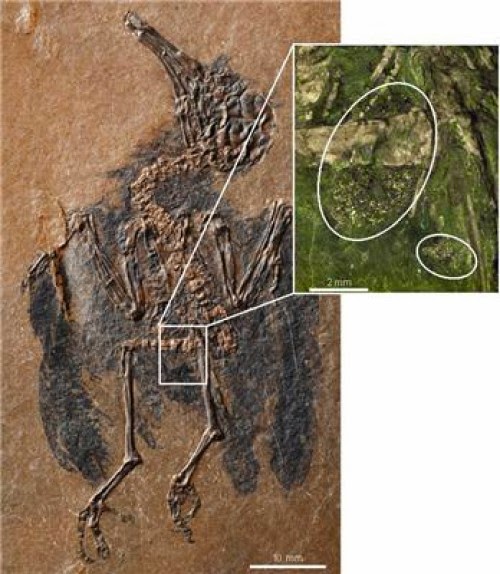
Scientists of the Senckenberg Research Institute in Frankfurt have described the oldest known fossil of a pollinating bird. The well-preserved stomach contents contained pollen from various flowering plants. This indicates that the relationship between birds and flowers dates back at least 47 million years. The fossil comes from the well-known fossil site “Messel Pit.” The study was published today in the scientific journal Biology Letters.
They fly from flower to flower, and with their long, slender bills they transfer the pollen required for the plants’ reproduction. Particularly in the tropics and subtropics, birds, besides insects, serve as the most important pollinators.
“While this process is well known and understood in the present, geological history has offered very little evidence of pollination through birds,” says Dr. Gerald Mayr, head of the Ornithological Section at the Senckenberg Research Institute in Frankfurt. He adds, “there have been occasional hints, such as characteristic bill shapes, that nectarivorous birds occurred in the past, but, so far, there existed no conclusive evidence.”
Now, however, the ornithologist from Frankfurt and his colleague, paleobotanist Dr. Volker Wilde, have found this evidence. In the well-preserved stomach contents of a fossil bird unearthed in the Messel Pit, the scientists discovered fossilized pollen grains.
“This is another discovery that underlines the unique significance of the Messel fossil site,” exclaims a delighted Dr. Wilde. “Not only does the presence of pollen offer direct evidence of the bird’s feeding habits, but it shows that birds already visited flowers as long as 47 million years ago!”
Fossil evidence for the existence of pollinating insects dates back to the Cretaceous period. Until now, however, there had been no information at what time pollination through vertebrates, and birds in particular, came into existence. To date, the oldest indication of an avian pollinator came from the early Oligocene, about 30 million years ago. “But this hummingbird fossil only offers indirect evidence of the existence of nectarivorous birds,” explains Mayr. “Thanks to the excellent state of preservation of the Messel bird, we were able to identify two different types of pollen, which is the first conclusive proof of nectarivory.”
Large numbers of differently sized pollen grains were found in the stomach contents of the completely preserved avian fossil. “Along with the bird’s skeletal anatomy, this indicates that we indeed have the fossil of a nectarivorous bird” explains Wilde.
And the spectacular discovery also suggests another conclusion: If a pollinating bird lived as much as 47 million years ago, it must be assumed that some representatives of the flora at that time had already adapted to this mode of pollination.
“To date, there are no fossil plants from this geological era that offer proof of the existence of ornithophily — i.e., the pollination of flowers through birds,” adds paleobotanist Wilde.
“However, the characteristic traits of bird-pollinated plants, such as red flowers or a lack of scent, do not fossilize,” elaborates Mayr. This lends an even greater importance to discoveries such as the Messel bird to understand the interactions between birds and flowers through geological time.
Note : The above story is based on materials provided by Senckenberg Research Institute and Natural History Museum.